By Ivy Geary, Eagle Journalism
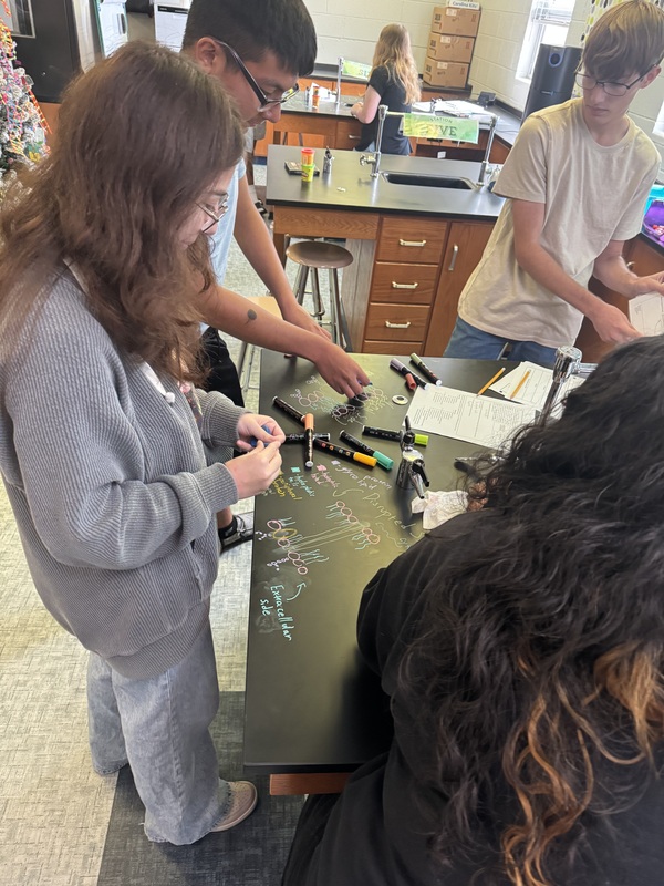
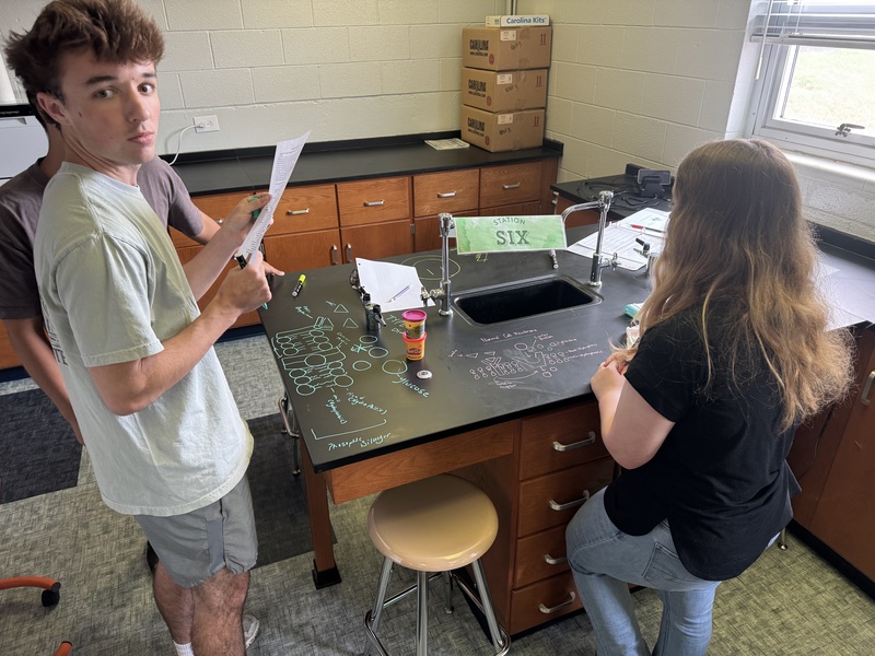
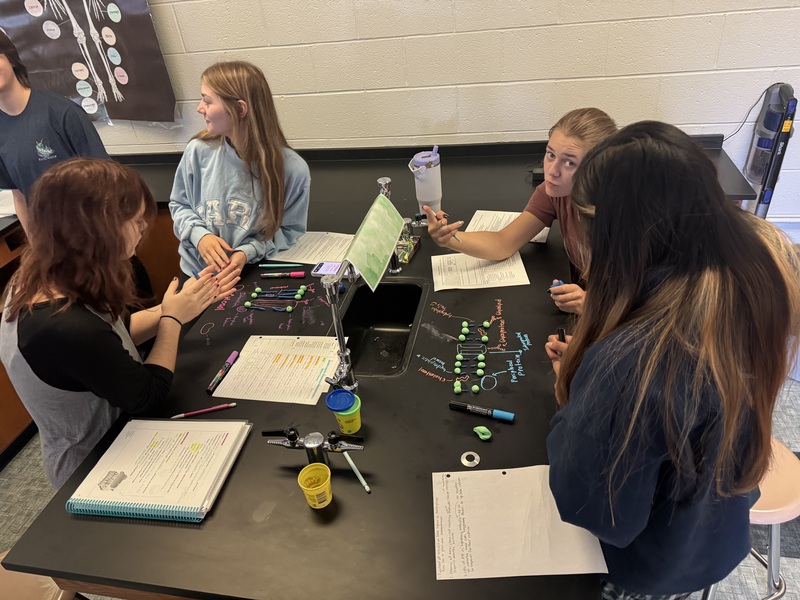
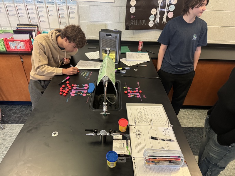
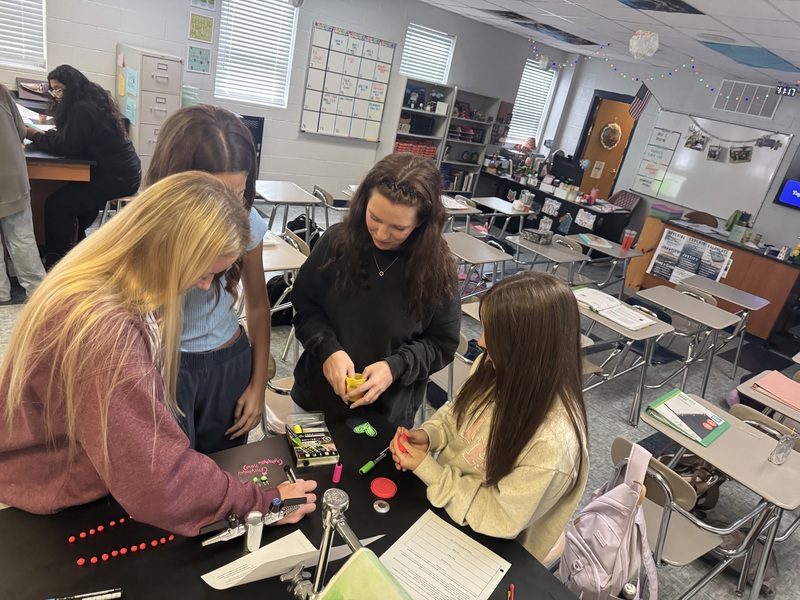
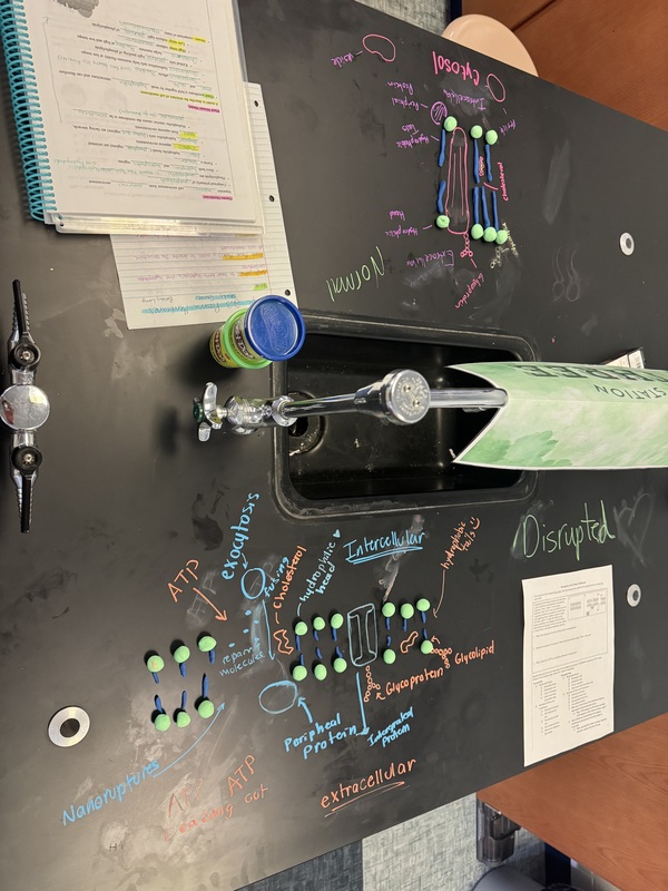
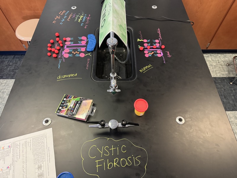
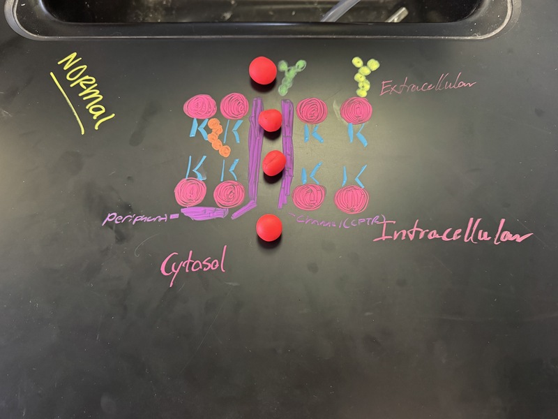
Recently Ms. Boyter’s AP Biology class used chalk markers and Playdoh to model what a cell membrane looks like before and after different diseases and disorders. By modeling the membrane not just after but before it is disrupted, students learn about the structure of the cell membrane and how each component works. Having a visual representation also helps students understand the process of cellular dysfunction.
The class then did a gallery walk and studied how each others’ abnormal cell membranes were disrupted. By doing a gallery walk and studying visual models of cell membranes normal and abnormal, they understand the anatomy of the cell better and understand how the defects connect to disease that has an impact on the world.